
Figure 4. CD276 blockade suppresses HNSCC growth and eliminates cancer stem cell in HNSCC.
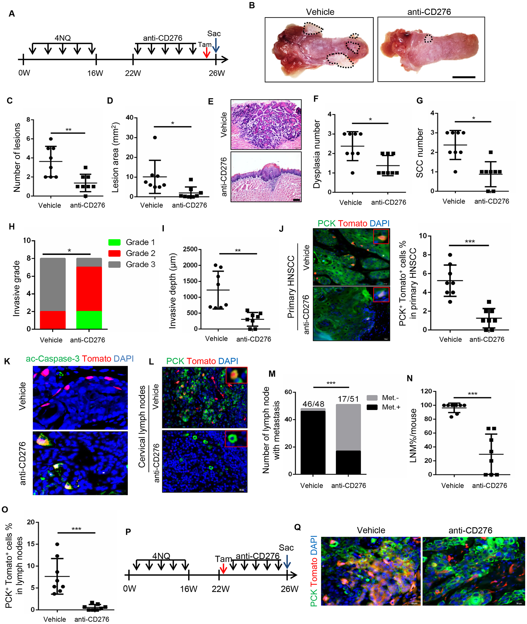
(A) Experimental design for the anti-CD276 treatment and lineage tracing of CSCs of HNSCC in Bmi1CreER;RosatdTomato mice.
(B) Representative image of tongue lesion 26 week in 4NQO-induced BmiCreER;RosatdTomato mice treated with anti-CD276 antibodies or vehicle with isotype IgG (vehicle). Scale bar, 3mm
(C) Quantification of lesion numbers visible in the mouse tongues. Values are mean ± SD from the pool of two independent experiments, n = 8, **p<0.01 by Student’s t test.
(D) Quantification of lesion areas visible in the mouse tongues. Values are mean ± SD from the pool of two independent experiments, n=8, *p<0.05 by Student’s t test.
(E) H&E staining of HNSCC in 4NQO-induced Bmi1CreER;RosatdTomato mice treated with anti-CD276 and vehicle with isotype IgG. Scale bar, 200μm
(F) Quantification of microscopic dysplasia numbers in mouse tongues. Values are mean ± SD from the pool of two independent experiments (n=8). *p<0.05 by Student’s t test.
(G) Quantification of microscopic SCC numbers in mouse tongues. Values are mean ± SD from the pool of two independent experiments (n=8). *p<0.05 by Student’s t test.
(H) Quantification of HNSCC invasive grades, n = 8, *p<0.05 by Cochran-Armitage test.
(I) Quantification of HNSCC invasive depths, n = 8, **p<0.01 by Student’s t test.
(J) Representative image and quantification of PCK+Tomato+ CSCs in primary HNSCC from mice treated with anti-CD276 or vehicle control with isotype IgG (n=8). Values are mean ± SD from the pool of two independent experiments (n=8). ***p<0.001 by Student’s t test. Scale bar, 20μm.
(K) Immunostaining of active caspase-3 (ac-Casp-3) in Tomato+ CSCs. Scale bar, 20μm.
(L) Representative image of PCK+ cancer cells and PCK+Tomato+ CSCs in cervical lymph nodes from mice treated with anti-CD276 antibodies. Scale bar, 20μm.
(M) Number of cervical lymph nodes with metastasis upon anti-CD276 treatment. ***p<0.001 by Fisher Chi square test. Met.−, without lymph node metastasis; Met.+, with lymph node metastasis.
(N) Percentage of lymph node metastasis in mice upon anti-CD276 treatment. ***p<0.001 by Student’s t test.
(O) Quantification of PCK+Tomato+ CSCs in cervical lymph node from mice treated with anti-CD276. ***p<0.001 by Student’s t test.
(P) Experimental design used to trace Bmi1+-derived tumor tissues in Bmi1CreER;RosatdTomato mice.
(Q) PCK immunostaining and Bmi1+-derived tumor tissues in BmiCreER;RosatdTomato mice upon anti-CD276 treatment. Scale bar, 20μm