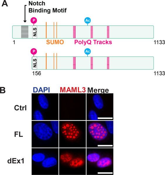
Figure 2. MAML3 constructs and in vitro cellular localization.
A, Diagram of MAML3 constructs showing predicted sites for posttranslational modifications, motifs, and nuclear localization signals. B, Immunofluorescence of SK-N-SH cells transfected with either FL or dEx1 MAML3 and stained for MAML3 show the nuclear localization. FL MAML3 showed punctate areas in the nucleus, whereas dEx1 MAML3 showed a more diffuse staining pattern. No endogenous MAML3 could be detected in transfected controls. (100x/1.30 oil Nikon Plan Fluor Objective 1.5x Zoom, AlexaFluor546, Scale bar represents 10 μm)

