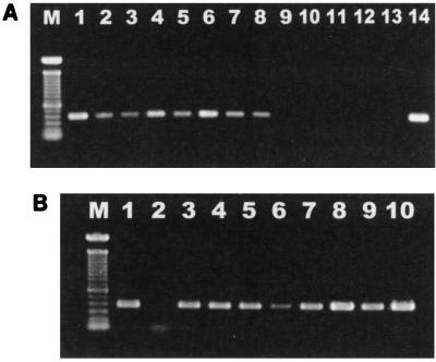
FIG. 2.
(A) Electrophoresis of DNA isolated from colonic biopsy samples, amplified by PCR with Helicobacter genus-specific primers, and run on a 1% agarose gel. Lane M, 100-bp DNA ladder; lane 1, MIT 97-6194-6; lane 2, MIT R97-6194-7; lane 3, MIT 97-6194-5; lane 4, MIT 97-6194-4; lane 5, MIT 97-6194-3; lane 6, MIT R97-6837; lane 7, MIT R97-6834; lane 8, MIT 97-6196-8; lane 9, MIT R97-6841; lane 10, MIT R97-6835; lane 11, MIT R97-6832; lane 12, MIT R97-6836; lane 13, blank; lane 14, positive control. (B) Electrophoresis of DNA isolated from fecal cultures, amplified by PCR with Helicobacter genus-specific primers, and run on a 1% agarose gel. The faint band in lane 2 at approximately 100 bases represents the PCR primers. Lane M, 100-bp DNA ladder; lane 1, positive control; lane 2, blank; lane 3, MIT 97-6194-4; lane 4, MIT 97-6194-3; lane 5, MIT 97-6194-5; lane 6, MIT R97-6834; lane 7, MIT R97-6841; lane 8, MIT R97-6837; lane 9, MIT R97-6840; lane 10, MIT R97-6842.

