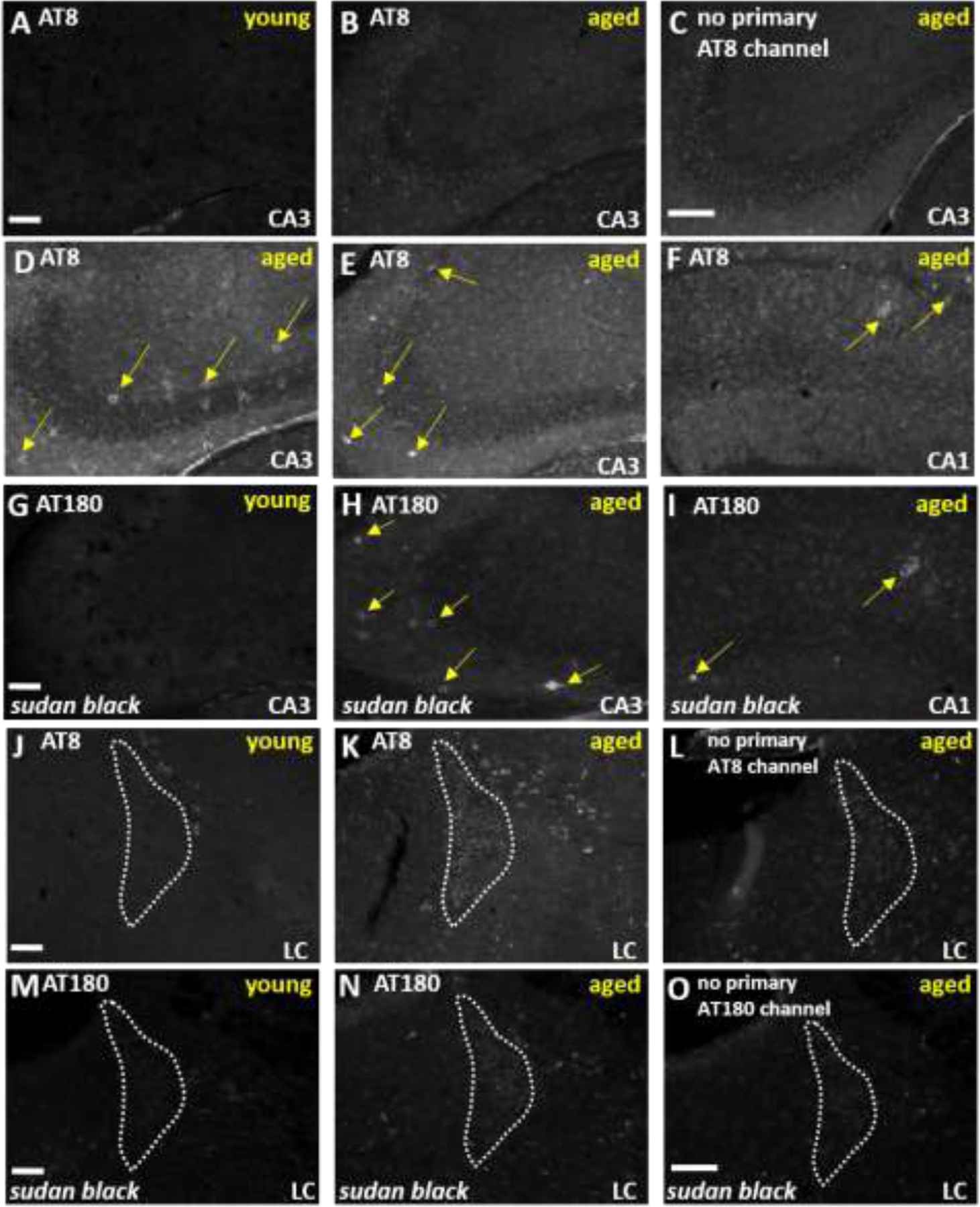
Figure 7.

Aged mice have evidence for AT8 and AT180-immunoreactivity in the hippocampus and locus coeruleus in addition to a distinctive age-related autofluorescence. Photomicrographs of the CA3 region of the hippocampus show representative AT8-immunoreactivity in A,B) young and aged mice as compared to C) no primary control in aged mice. D-F) Distinctive AT8-immunoreactivity is depicted in CA3 and CA1 of two independent aged mice (2 out of 10 mice had this type of staining). G-I) AT180 immunoreactivity is depicted in young and aged tissue treated with Sudan Black in order to reduce non-specific background staining. Panels H and I show a similar immunoreactivity pattern with AT180 as observed with the AT8 antibody in the same subjects as panels D-F. Photomicrographs of the locus coeruleus at −5.62 mm Bregma show representative J,K) AT8-immunoreactivity in young versus aged mice alongside L) a no primary control in aged mice. M,N) AT180 immunoreactivity is depicted in young and aged tissue treated with Sudan Black. O) No antibody control for the AT180 immunostaining conditions. White scale bars equal 100 micrometers.
