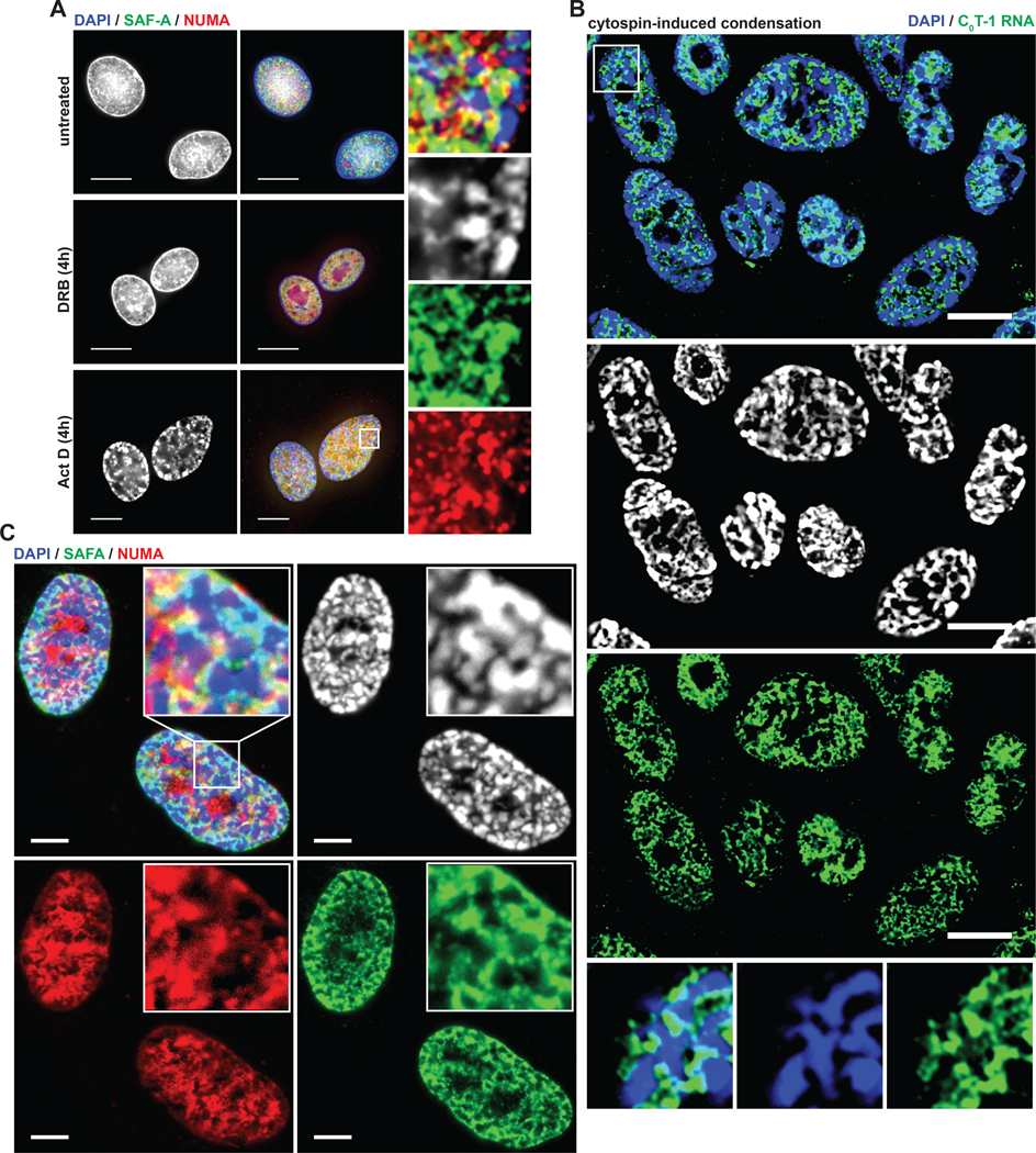
Figure 6. Euchromatin-associated scaffold proteins are depleted from experimentally condensed chromatin.
(A) IF analysis of cells after treatment with transcriptional inhibitors as indicated. Higher magnification of the indicated region on right. Arrows indicate high DAPI intensity bodies depleted of SAF-A and NUMA. Scale bars 10 μm. (B,C) FISH and IF analysis of cells after high-speed cytocentrifugation. All cells analyzed (n = 200) had many visibly condensed chromatin bodies following high-speed cytospin attachment to coverslips (2000 RPM, 10 minutes) compared to 6% (n = 200) of cells attached to coverslips using a standard cytospin procedure (800 RPM, 3 minutes). High DAPI intensity bodies are indicated by arrows at higher magnification.

