
Figure 3:
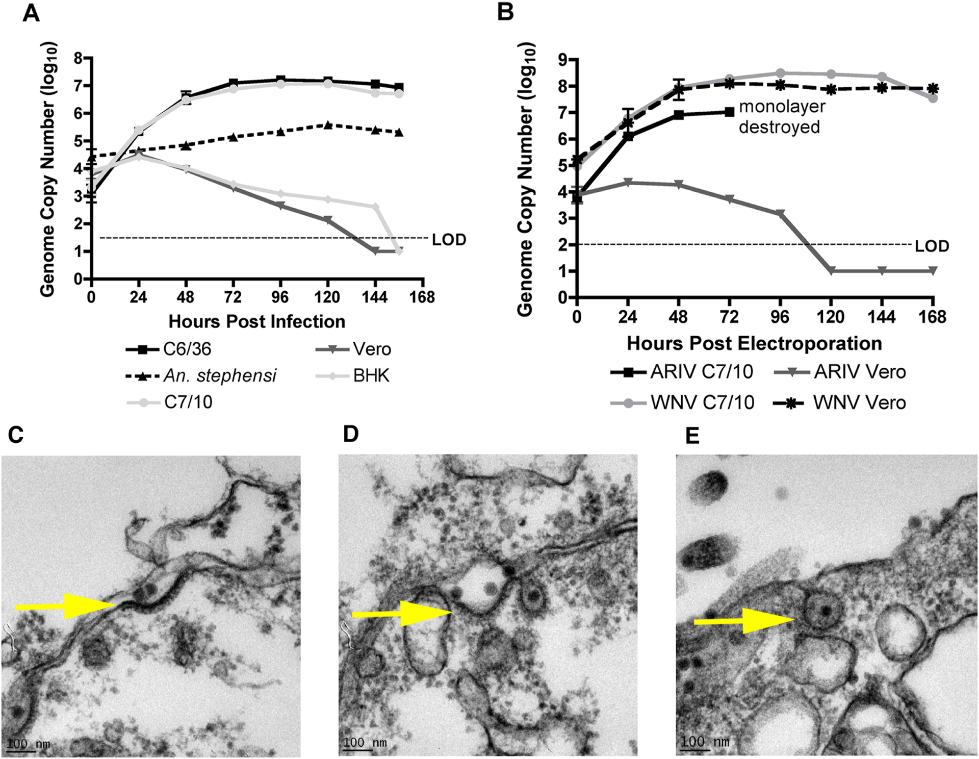
Replication kinetics of ARPV in representative mosquito and vertebrate cell lines post-infection (A) and post-electroporation (B). Monolayers were inoculated with an MOI of ~0.1. Data points represent the mean number of genome copies/ml for duplicate infections titrated in triplicate using qPCR tests in (3A) and triplicate infections titrated in triplicate in (3B). Error bars indicate the standard deviation of the mean. Transmission electron micrographs of ARPV-infected Vero cells show that ARPV enters by clathrin-mediated endocytosis by 15 sec (c), 1 min (d), and 5 mins (e) post-infection. An ARPV virion is localized at the surface of the cellular plasma membrane (c). A clathrin-coated pit is formed, and endocytosis is initialized after viral protein-host cell receptor binding (d) prior to ARPV entry via endocytosis (e). Scale bar (100 nm) is shown in the bottom left of each micrograph.