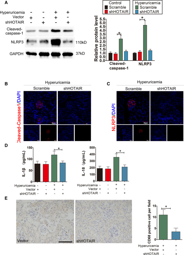
FIGURE 8.

Knockdown of HOTAIR ameliorates renal inflammation in HUA mice. (A) The protein levels of caspase‐1, NLRP3, GSDMD‐N and GSDMD‐FL, as measured by Western blot analysis; quantification normalized to GAPDH in renal tissue from hyperuricaemia mice after shRNA treatment. The protein levels of caspase‐1, NLRP decreased remarkably in hyperuricaemia+shHOTAIR group; *p < 0.05 compared to the hyperuricaemia +vector group; n = 6 in each group. (B,C) Immunofluorescence images showing the expression of caspase‐1, NLRP3 in renal tissue from hyperuricaemia mice after shRNA treatment. Tissue immunofluorescence showed that the fluorescence intensity of glomerular NLRP3 and caspase‐1 in the shRNA‐treated group was significantly lower than that in the hyperuricaemia+shHOTAIR group; *p < 0.05 compared to the control group; n = 6 in each group (scale bar, 50 μm; magnification, 400×); blue: nuclear staining (DAPI), red: caspase‐1 and NLRP3 staining. (D) The serum levels of IL‐1β and IL‐18, measured by ELISA. The serum levels of IL‐1β and IL‐18 decreased remarkably in the hyperuricaemia+shHOTAIR group; *p < 0.05 compared to the in the hyperuricaemia+Vector group; n = 6 in each group. (E) CD68 immunohistochemistry of the renal interstitium and glomerular mesangial area in the hyperuricaemia+vector group and hyperuricaemia+shHOTAIR group. Immunohistochemistry of CD68 in renal tissue from hyperuricaemia mice after shRNA treatment. Compared with that in the hyperuricaemia+Vector group, the down‐regulation of HOTAIR by shHOTAIR significantly reduced the infiltration of positive CD68 macrophages in the renal interstitium and glomerular Mesangial area in hyperuricaemia+shHOTAIR group; ∗p < 0.05 compared to hyperuricaemia+vector group; n = 6 in each group (scale bar, 100 μm; magnification, 400×)
