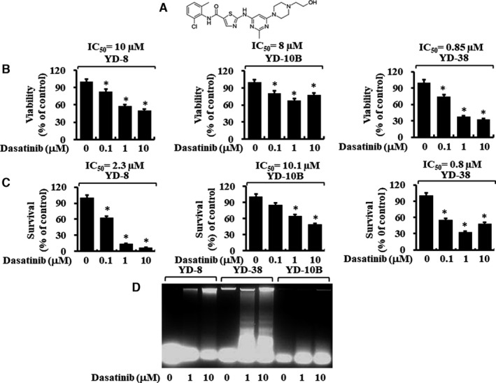
FIGURE 1.

Effects of dasatinib on cell viability, cell survival and apoptosis (DNA fragmentation) on three different human oral cancer cells. A, The chemical structure of dasatinib. B‐C, YD‐8, YD‐38 and YD‐10B cells were treated with dasatinib (0, 0.1, 1 and 10 μM) for 24 h, followed by measurement of cell viability by MTS assay (B) and cell survival by cell count analysis (C). Data are mean ±SE of three independent experiments. *P < .05 compared with the values of control (no dasatinib). D, YD‐8, YD‐38 and YD‐10B cells were treated with dasatinib (0, 1 and 10 μM) for 8 h. Extra‐nuclear fragmented DNA was extracted and analysed on a 1.7% agarose gel
