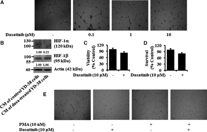
FIGURE 8.

Effects of conditioned media (CM) from control‐ or dasatinib‐treated YD‐38 cells on tube formation, viability, survival, and HIF‐1α protein expression of HUVEC. (A‐B) YD‐38 cells were treated without or with dasatinib at the indicated doses for 24 h. The conditioned media (CM) was then collected and applied to HUVEC cultured in Matrigel‐coated plates for an additional 16 h. Changes in cell morphology (HUVEC tube formation) were captured using an inverted microscope (A). Whole‐cell lysates were prepared and analysed by Western blotting (B). (C‐D) HUVECs were treated without or with dasatinib (10 μM) for 16 h, followed by measurement of cell viability and cell survival by MTS (C) and cell count (D) assays, respectively. Data are the means ±SE of three independent experiments. (E) YD‐38 cells were treated without or with PMA (10 nM), a known inducer of HUVEC tube formation, in the absence or presence of dasatinib (10 µM) for 8 h. The conditioned media (CM) was then collected and applied to HUVEC cultured in Matrigel‐coated plates for an additional 16 h. Changes in cell morphology (HUVEC tube formation) were captured using an inverted microscope
