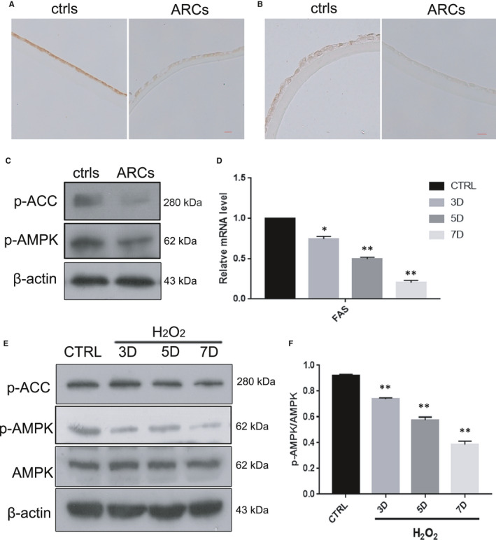
FIGURE 3.

The AMPK pathway was inactivated in the human lens epithelium of ARCs and in HLE‐B3 cells with oxidative stress‐induced senescence (A). Representative images from IHC assays against p‐AMPKα (Thr172) in the controls (left) and ARCs (right). (B). Representative images from IHC assays against p‐ACC (ser79) in the controls (left) and ARCs (right). (C). Representative images from Western blot analysis of p‐AMPKα (Thr172), p‐ACC (ser79) and β‐actin in the controls (left) and ARCs (right). (D). Relative fold‐changes in the mRNA levels of the genes encoding FAS, as determined by qRT‐PCR. (E). Western blot analysis of AMPKα, p‐AMPKα (Thr172), p‐ACC (ser79) and β‐actin in HLE‐B3 cells treated with H2O2 (150 μM) for 3–7 days. (F). Relative fold‐changes in the protein levels of p‐AMPKα/AMPKα as described in E. Data were shown as mean ± SD and are representative of 3 independent experiments. *p < 0.05; **p < 0.01 compared to the control (CTRL). The bar represents 50 um
