Figure 5.
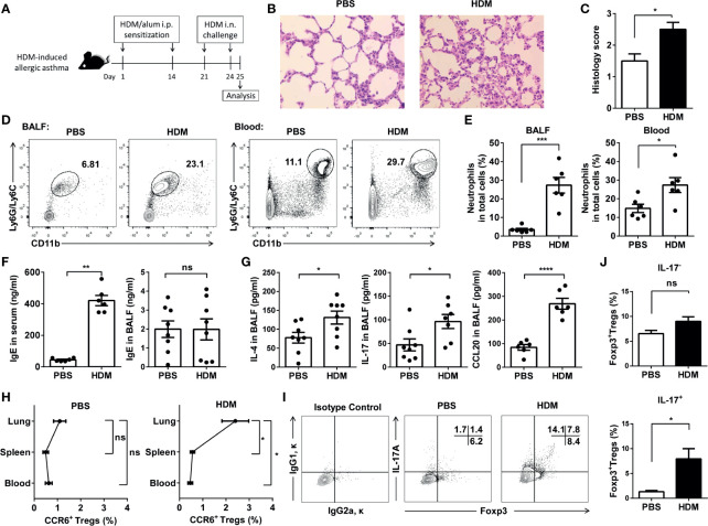
CCR6+Treg cells accumulated in lungs during HDM-induced allergic airway inflammation. (A) Experimental scheme. Mice were sensitized and challenged as described. (B) H&E staining of the lung tissues. (C) Determination of pathology score as described in Materials and Methods (n = 5 mice). (D, E) Neutrophils in the BALF and blood from asthmatic (n = 6) and control mice (n = 6) were analyzed by FACS. Neutrophils were identified as CD11b+Ly6G/Ly6C+ cells. (F) Determination of IgE secretion in serum and BALF using ELISA. (G) Determination of IL-4, IL-17 and CCL20 levels in BALF. (H) FACS analysis of CCR6+ Treg cells in the lung, spleen, and blood from HDM-induced asthmatic and control mice. Identification of CCR6+ Treg cells as CD4+CCR6+CD25+CD127- cells. (I, J) FACS analysis of IL-17-Foxp3+ Treg cells and IL-17+Foxp3+ Treg cells in BALF from HDM-induced asthmatic (n = 5) and control mice (n = 5). Data are representative of three independent experiments with ≥ four mice per group. *P < 0.05, **P < 0.01, ***P < 0.001; ns, not significant.

