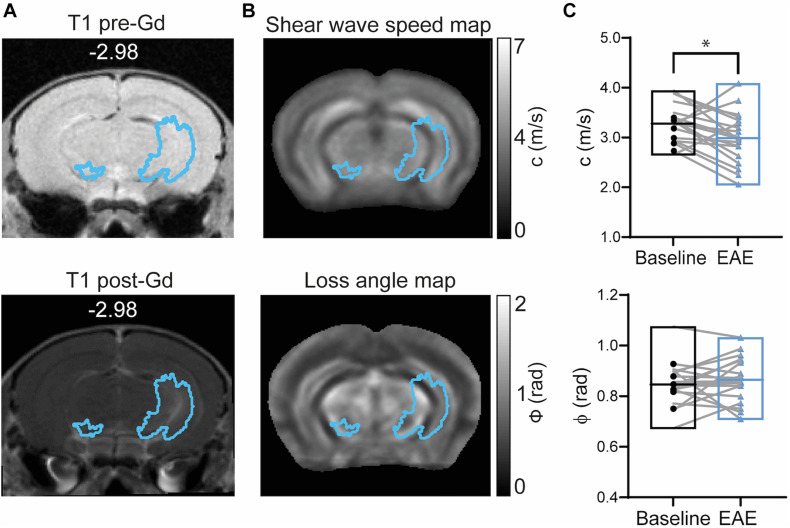
FIGURE 5.
Brain mechanical properties in Gd-enhanced lesions during EAE. (A) Representative T1-weighted image acquired before (left) and after GBCA (right) injection (corresponding GBCA mask overlaid) shows Gd-enhancing lesions near the left hippocampal artery. (B) GBCA mask overlaid on averaged MRE maps of stiffness (shear wave speed) and fluidity (loss angle). (C) Stiffness represented by shear wave speed (c) was reduced during EAE (p = 0.0183; paired t-test) whereas tissue fluidity (ϕ) was unchanged (p = 0.3683; paired t test). n = 19; mean, min/max; *<0.05. Of note, ventricles are excluded from our analysis since MRE cannot be performed in fluid compartments.

