
Figure 1.
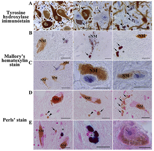
NM-containing neurons and eNM in human SN pars compacta. A) Tyrosine hydroxylase immunostaining counterstained with hematoxylin. B,C) Mallory’s hematoxylin staining. D,E) Perls’ staining. A) Most neurons in human SN pars compacta have NM in the cytoplasm and are either intensely TH-immunoreactive (arrow 1) or moderate/weak TH-immunoreactive (arrow 2); some of neurons in SN pars compacta are NM-containing and TH-immunonegative (arrow 3); single SN pars compacta neurons are TH-immunonegative and do not contain NM (arrow 4). The majority of human SN pars compacta samples analyzed contain accumulations of eNM (arrowhead). B,C) Intracellular NM does not react with Mallory's hematoxylin, remaining golden brown; dark blue are small, fine-grained clusters in the neuropil (double arrow), destroyed neurons (extracellular neuromelanin,- eNM), and macrophages (arrow); in 2 cases excluded from the initial samples due to prolonged illness of subjects, we found intense blue staining of the nucleus and intranuclear structures in some of the dopaminergic neurons in SN pars compacta (C, line). D,E) After Pearls' reaction for iron, no staining of intracellular NM and eNM (arrowhead) was observed; some parenchymal macrophages contained golden brown granules in the cytoplasm (arrow); some perivascular macrophages expressed dark blue staining of the cytoplasm due to the presence of the pigment hemosiderin, which contains iron oxide; the asterisk is the lumen of the blood vessel; as with the use of Mallory's hematoxylin, in 2 cases, the nucleolus of some SN pars compacta neurons was stained blue (line), which indicates the accumulation of non-heme iron in the nucleoli of these cells. Scale bar: 20 μm.