
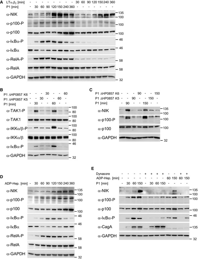
Figure 1. H. pylori‐induced classical and alternative NF‐κB pathways depend on ADP‐heptose.
-
AAGS cells were infected with H. pylori or treated with 20 ng/ml LTα1β2 for the indicated times. Total cell lysates were analyzed by immunoblotting using the indicated antibodies.
-
B, CAGS cells were infected with H. pylori P1 wild‐type or isogenic ΔHP0857 (mutated in the gmhA gene) strains. Total cell lysates were analyzed by immunoblotting using the indicated antibodies.
-
DAGS cells were treated with 200 nM synthetic ADP‐heptose for the indicated times. Total cell lysates were analyzed by immunoblotting using the indicated antibodies.
-
EAGS cells were pre‐incubated with 100 μM dynamin GTPase inhibitor Dynasore for 60 min followed by H. pylori infection or addition of ADP‐heptose (200 nM). Total cell lysates were analyzed by immunoblotting using the indicated antibodies. CagA demonstrated a similar infection rate.
Data information: Data shown are representative for at least two independent experiments. GAPDH served as loading control.
Source data are available online for this figure.