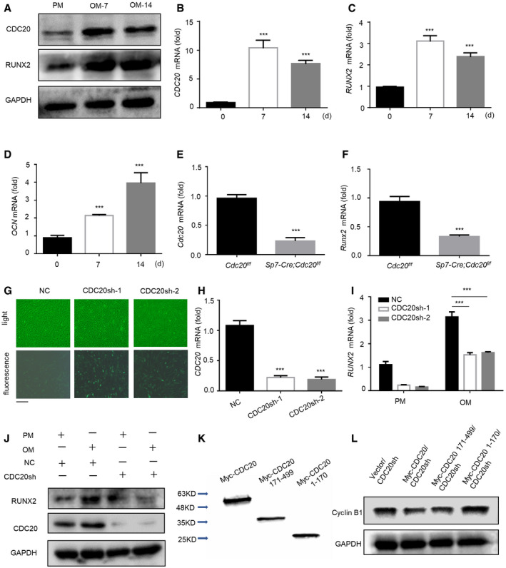
Figure EV2. CDC20 modulates osteogenic differentiation of BMSCs, related to Fig 2 .

-
A–DWestern blot analyses (A) and qRT–PCR (B‐D) of the expression of CDC20 and osteogenic marker RUNX2, OCN. Cells were cultured in osteogenic medium for 7 and 14 days (n = 6).
-
E, FThe knockout efficiency of Cdc20 (E) and the expression of osteogenic marker Runx2 (F) in BMSCs of Sp7‐Cre;Cdc20f / f and Cdc20f / f mice determined by qRT–PCR (n = 5).
-
GRepresentative images of light and fluorescence of lentivirus infected NC and CDC20sh hBMSCs. Scale bar: 500 μm.
-
HThe knockdown efficiency of CDC20 in NC and CDC20sh hBMSCs determined by qRT–PCR (n = 5).
-
I, JThe expression of RUNX2 in NC and CDC20sh hBMSCs after 7 days osteogenic differentiation determined by qRT–PCR (I) and Western blot analyses (J) (n = 5).
-
KWestern blot analyses of Myc‐CDC20, Myc‐CDC20 171–499 fragment (containing WD40 domain), Myc‐CDC20 1–170 fragment (lacking WD40 domain) plasmids expression in HEK293T cells.
-
LWestern blot analyses of the degradation of the substrate Cyclin B1 under the overexpression of truncated fragments of CDC20.
Data information: Data are displayed as mean ± SD and show one representative of n ≥ 3 independent experiments with three biological replicates. Statistical significance was calculated by a two‐tailed unpaired Student’s t‐test or one‐way ANOVA followed by a Tukey’s post hoc test and defined as ***P < 0.001.
