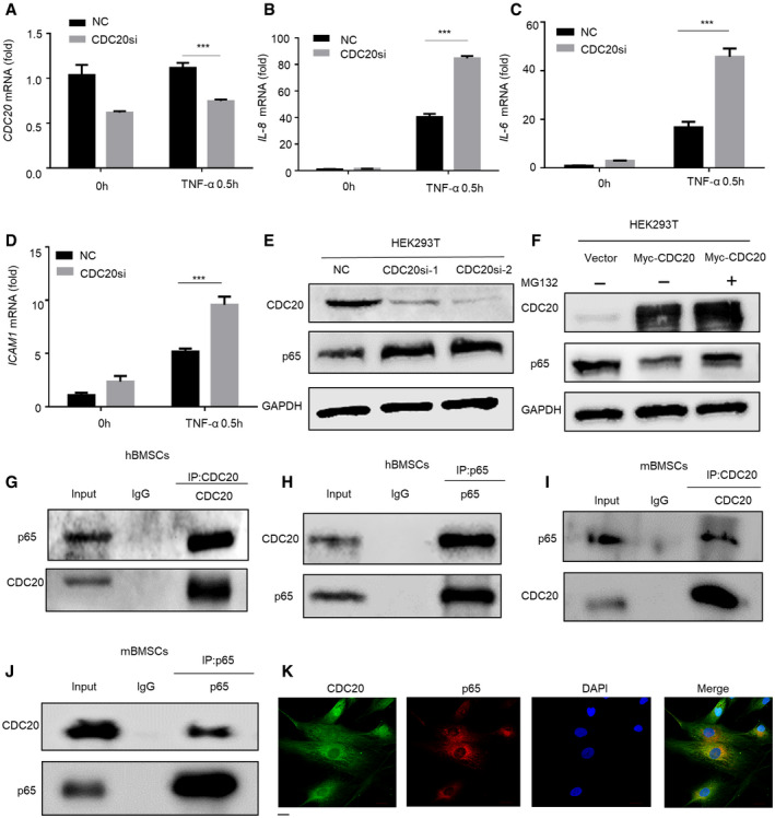
Figure EV3. CDC20 induces proteasome‐dependent degradation of p65 and CDC20 interacts with p65, related to Figs 3 and 4 .

-
A–DThe expression of CDC20 (A) and NF‐κB pathway downstream genes IL‐8, IL‐6, and ICAM1 (B‐D) of NC and CDC20si HEK293T cells after TNF‐α stimulation determined by qRT–PCR (n = 6).
-
EWestern blot analyses of the degradation of endogenous p65 protein in NC and CDC20si HEK293T cells.
-
FWestern blot analyses of the degradation of p65 protein after the overexpression of Myc‐CDC20. HEK293T cells were transfected with Vector and Myc‐CDC20 plasmids for 36 h, cells were treated with or without 10 μM MG132 (the proteasome inhibitor) for 6 h before collected.
-
G, HCo‐immunoprecipitation of endogenous CDC20 with endogenous p65 in hBMSCs.
-
I, JCo‐immunoprecipitation of endogenous CDC20 with endogenous p65 in mBMSCs.
-
KThe co‐localization of CDC20 and p65 in hBMSCs. Scale bar: 20 μm.
Data information: Data are displayed as mean ± SD and show one representative of n ≥ 3 independent experiments with three biological replicates. Statistical significance was calculated by one‐way ANOVA followed by a Tukey’s post hoc test and defined as ***P < 0.001.
