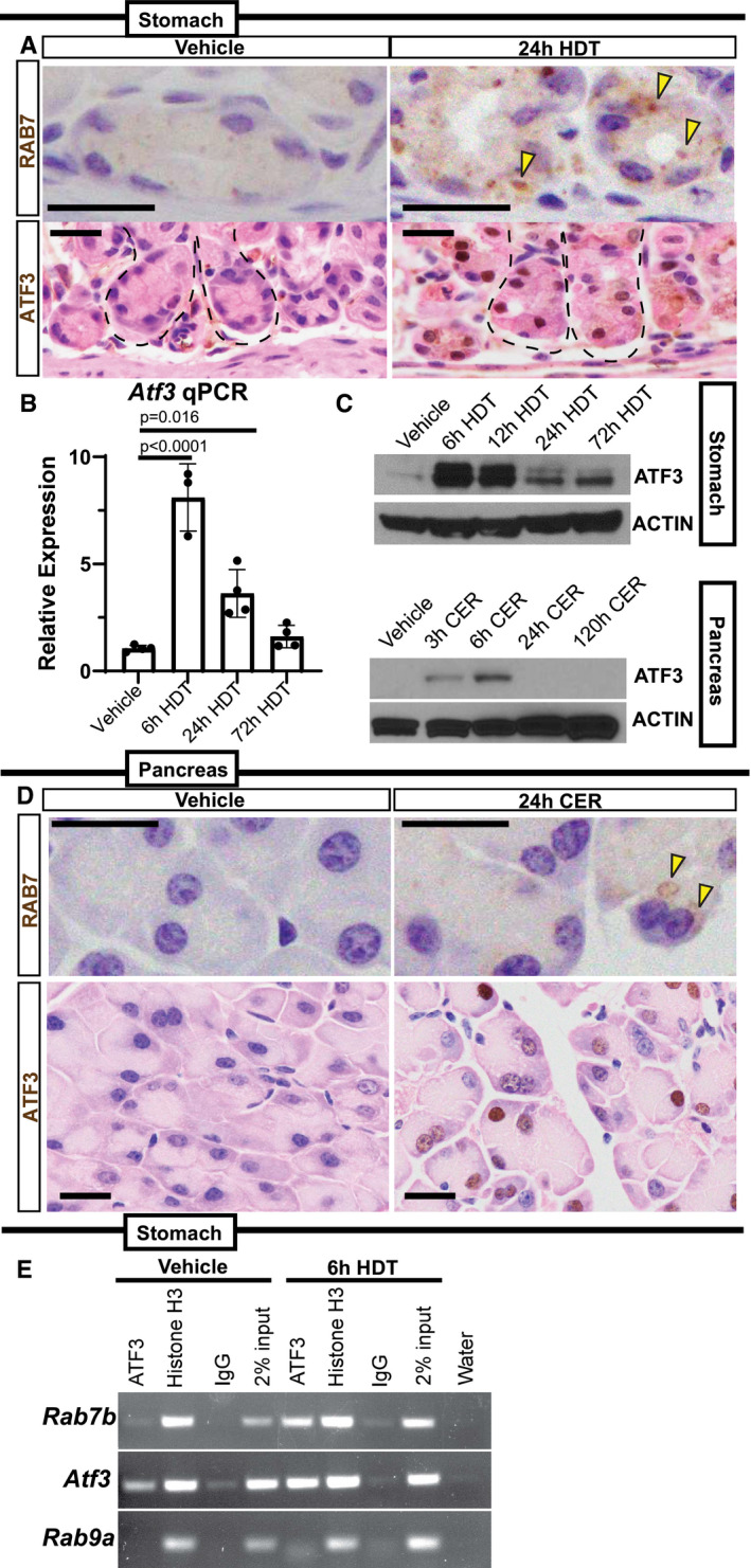
Top—Representative RAB7 expression in base of stomach units (ZCs) in vehicle or 24‐h HDT by immunohistochemistry. Arrowheads point to RAB7 vesicle aggregates. Scale bar 20 µm. Bottom—Representative ATF3 expression in base of stomach units (ZCs) by immunohistochemistry. ATF3 appears in nuclei following 24‐h high‐dose tamoxifen (HDT). Scale bar 20 µm. Stomach unit base outlined by dashed black line.
Atf3 mRNA expression by qRT–PCR in vehicle, 6, 24, and 72‐h HDT treatment. Each data point represents the mean of triplicate technical replicates from a single mouse, n = 3–4 mice in independent experiments. Significance determined by one‐way ANOVA, Tukey. Error bars denote standard deviation.
Western blot of ATF3 expression over HDT and CER time courses with β‐Actin loading control.
Top—Representative RAB7 staining in pancreatic acinar cells. Arrowheads indicate RAB7 vesicles. Scale bar 20 µm. Bottom—Representative ATF3 expression in pancreatic acinar cells. Nuclei express ATF3 after 24‐h cerulein (CER) injury. Scale bar 20 µm.
ChIP‐PCR on mouse stomach tissue at vehicle and 6‐hour HDT. Chromatin probed with ATF3, Histone H3 antibodies, or a Rabbit IgG control. Also included is the 2% chromatin input and water to control for DNA contamination. Rab7b amplicon is from a conserved, putative ATF3‐binding site in the first intron. Amplicon from Atf3 is a positive control from a previously characterized ATF3‐binding site; negative control is from a site in Rab9a lacking putative ATF3‐binding motifs.

