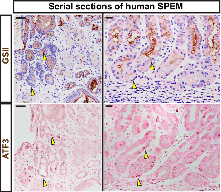
Figure 6. ATF3 is expressed in human SPEM.

Serial sections of human tissue. Representative immunohistochemistry of GSII (top; spasmolytic polypeptide‐expressing metaplasia [SPEM] marker and neck cells) counterstained with hematoxylin (blue) and eosin (pink). Representative staining of ATF3 (bottom) counterstained with eosin (pink). Tissue was taken from regions adjacent to gastric adenocarcinoma. Scale bars left—50 μm and right—20 μm. Arrowheads indicate strongly expressing ATF3 cells and the corresponding region in the same gland labeled with GSII in a serial tissue section. GSII at the base of gastric glands in the body of the stomach indicate transition to SPEM.
