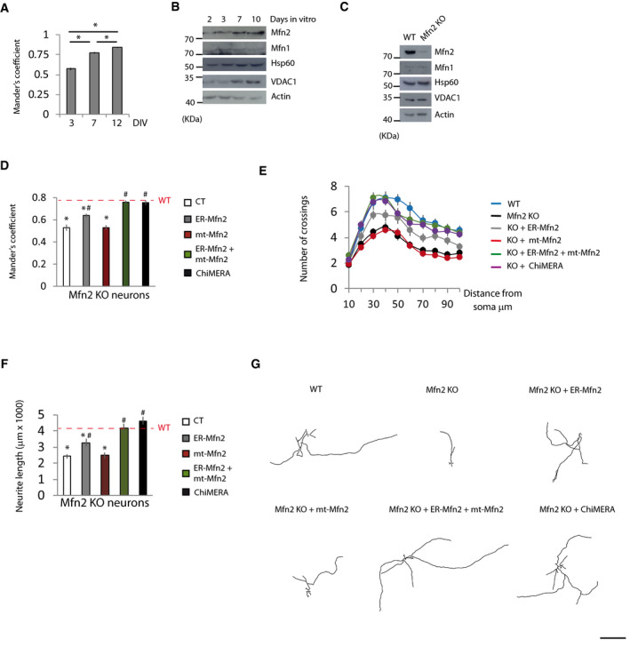
Figure 6. Mfn2‐mediated ER‐mitochondria tethering is necessary for neurite outgrowth.

-
APrimary cortical neurons were transfected 48 h before the indicated day in vitro (DIV) with mt‐RFP and ER‐GFP, fixed at the indicated DIV, and mitochondria‐ER colocalization was analyzed using Mander’s coefficient (n = 21 neurons were analyzed in three independent experiments). Data are presented as mean ± SEM.
-
BRepresentative Western blots of the indicated proteins during the neuronal maturation in vitro (n = 3 independent experiments).
-
CRepresentative Western blots of the indicated proteins of WT and Mfn2 KO neurons at DIV7 (n = 3 independent experiments).
-
DPrimary neurons of tamoxifen‐inducible Mfn2 KO mice were transfected at DIV3 with ER‐GFP, mt‐RFP plus the indicated plasmids. Tamoxifen‐treated (for 72 h) and vehicle‐treated neurons (WT; red dashed line) were fixed, and mitochondria‐ER colocalization was analyzed using Mander’s coefficient (n = 15 neurons analyzed in three independent experiments). Data are presented as mean ± SEM.
-
E–G(E) Sholl analysis and (F) neurite length measurement and representative tracings (G) of primary cortical neurons of WT and Mfn2 KO mice co‐transfected at DIV4 with GFP and the indicated plasmids. After 72 h, the neurons were fixed, immunofluorescence with anti‐GFP antibodies was performed and neurite length was measured (n = 30 neurons from three independent experiments). Data are presented as mean ± SEM.
Data information: *P < 0.05 versus WT; # P < 0.05 versus Mfn2 KO neurons, one‐way ANOVA followed by Tukey’s post hoc test. Scale bar: 500 µm.
