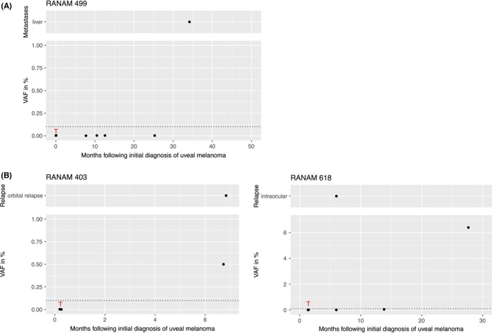
FIGURE 2.

VAFs of mutant GNAQ or GNA11 alleles in UM patients at different time points after the initial diagnosis of the primary tumor (Lower part). (A) Example of a metastatic patient who did not show an increase in ctDNA until the end of the study. (B) Two patients with intra‐ or extraocular recurrence. Red T: Time point of sampling of tumor tissue. Dotted line: level of detection at VAF = 0.1%. Upper part: location and time of clinical diagnosis of the recurrence (RAN403 after 6.8 months and RAN618 after 7 months). UM, uveal melanoma; VAF, variant allele fraction
