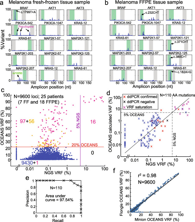
Fig. 5.
Validation of the 15-plex OCEANS melanoma panel on melanoma tissue samples. a Example results from a fresh-frozen melanoma tissue sample. The BRAF-V600E mutation was the only mutation called for this sample. b Example results from a formalin-fixed, paraffin-embedded (FFPE) tissue sample. 4 mutations were called in the AKT1, MAP2K1, MAP2K2, and NRAS genes. Other variants with ≥20% VRF that were not called by Clair are not labeled in the figure. All 4 of these mutations had confirmatory reads on a parallel NGS experiment, but only the NRAS c. 182A >G mutant had a NGS VRF of above 5%; see Additional file 1: Section S5 for details. c Summary of sequencing results for 25 clinical melanoma tissue samples (7 fresh/frozen, 18 FFPE). Input DNA quantities ranged from 10 ng and 50 ng (Additional file 2). The X-axis shows the VRF based on a standard NGS analysis, and the Y-axis shows the OCEANS VRF. The horizontal line shows the 20% VRF cutoff for OCEANS variant calls, and the vertical line shows the 5% VRF cutoff for NGS variant calls. The numbers in quadrants display the number of loci in each group; 97 of the 153 putative variants in the top-left quadrant also had a Clair score of above 180 (purple dots). Relative to the NGS results, the OCEANS panel had a sensitivity of 100% and a specificity of 99.0%, indicating a very a low false positivity rate. Importantly, we believe that many of the 97 NGS-negative and OCEANS-positive results were true mutations, and our ddPCR confirmation experiments support this hypothesis (Table 1). We did not observe any significant difference for fresh/frozen samples vs. FFPE samples (Additional file 1: Fig S12). d Comparison of OCEANS calculated VAF and NGS VRF. Original sample VAF was calculated for mutation calls from OCEANS VRF in panel (c) (Additional file 1: Section S4). Based on calibration experiments in Fig. 4 d, VAF quantitation dynamic range is relatively small due to VRF saturation. However, VAF estimation enables identification of high VAF mutations (>5%) to aid treatment decisions based on clinical diagnosis. Due to nanopore error rate, OCEANS %VRF >90% was considered as saturated and classified as high VAF mutation irrespective of the calculated VAF value. OCEANS identified all mutations with NGS VRF >5% as high VAF mutations. OCEANS called several low VAF mutations. Mutations confirmed by ddPCR are in red and a ddPCR negative mutation is indicated in black (Table 1). Note: 110 amino acid mutations (Additional file 2) from 114 mutated nucleotide loci that were called by Clair in panel (c) (pink dots) are plotted here. e Precision and recall for data in panel (d), based on changing the OCEANS calculated VAF cutoff. The area under the curve (AUC) is 97.54%. f High concordance of OCEANS results using Oxford Nanopore MinION vs. Flongle flow cells

