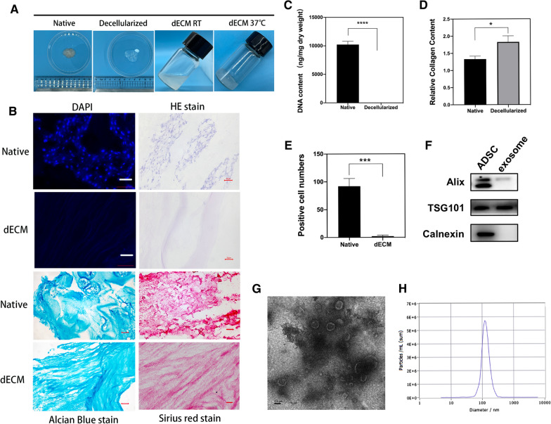
Fig. 1.
Preparation and identification of dECM and characterization of exosomes. A Images of nucleus pulposus tissue decellularization and preparation of hydrogels. B DAPI and H&E staining demonstrated decellularization; Alcian blue staining confirmed the retention of glycosaminoglycan; Sirius red staining confirmed retention of collagen fibers. Scale bar = 20 μm. C Quantitative determination of DNA content and D collagen retention. Data are expressed as the mean ± SD (n = 3). ****P < 0.0001, *P < 0.05. E Cell count in the fluorescence field (n = 3). F Exosome characterization of Alix, TSG101, and calnexin by Western blotting. G TEM analysis of exosomes. Scale bar = 0.1 μm. H NTA analysis of exosomes

