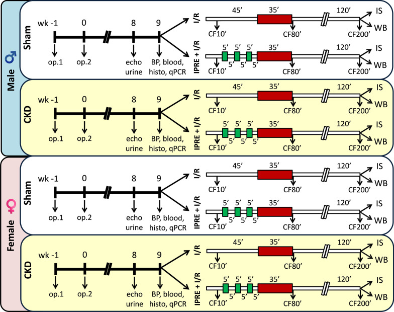
Fig. 1.
Protocol figure. Male and female Wistar rats underwent sham operation or 5/6 nephrectomy in 2 phases to induce chronic kidney disease (CKD). First, 2/3 of the left kidney was ligated and excised (Op1). One week later, the right kidney was removed (Op2). Corresponding time-matched sham operations were performed in the sham groups. At week 8, cardiac morphology and function were assessed by transthoracic echocardiography (echo) in a subgroup of animals. In this week, a subgroup of the animals was placed for 24 h into metabolic cages to collect urine for determination of urine creatinine and protein levels. At week 9, rats were anesthetized, and blood was collected from the thoracic aorta to measure serum urea and creatinine levels (blood). In this week, blood pressure was also measured in a subgroup of animals (BP). Hearts were then an isolated used for histology (histo) and qRT-PCR (qPCR) measurements or perfused according to Langendorff, the perfused hearts were subjected to global ischemia/reperfusion (I/R) with or without ischemic preconditioning protocol (IPRE, 3 × 5 min I/R cycles). Coronary flow (CF) was measured at the 10th, 80th, and 200th minutes of the perfusion protocol (CF10', CF80', and CF200', respectively). At the end of the perfusion protocol, hearts were collected for infarct size (IS) or Western blot (WB) measurements

