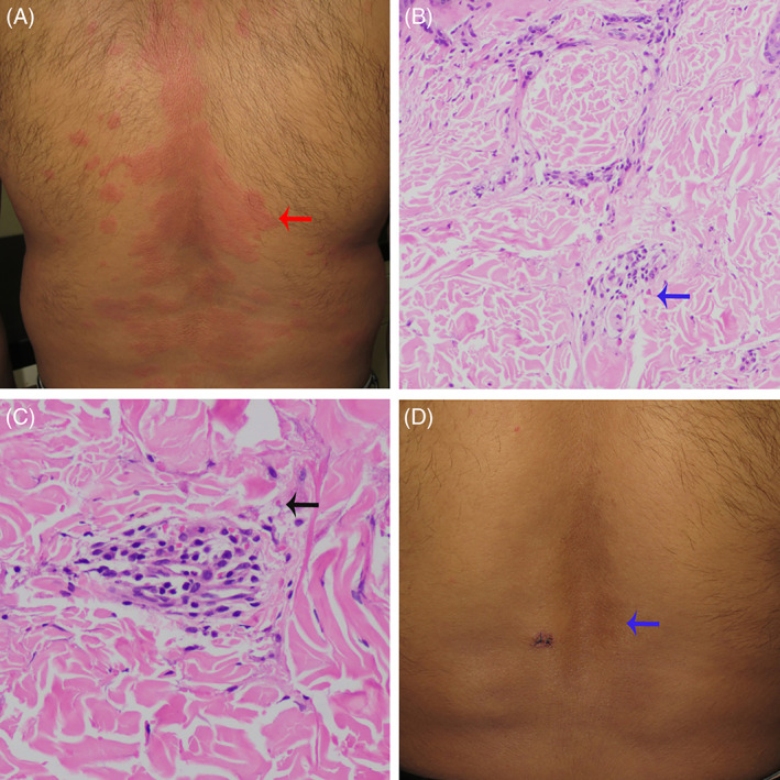
FIGURE 1.

(A) Multiple urticarial plaques (red arrow). (B) Histology shows mild perivascular infiltration comprising neutrophils, eosinophils (blue arrow), and lymphocytes (H & E, ×100). (C) Histology shows vessel wall disruption, erythrocyte extravasation (black arrow) (H & E, ×400). (D) Postinflammatory hyperpigmentation (blue arrow)
