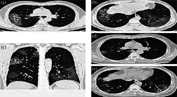
FIGURE 7.

A middle‐aged male with no comorbidities and h/o onset of high temperature and cough. Hemogram shows normal leukocytic ranges with reduced lymphocytes. Radiograph shows the appearance of patchy widespread infiltration in lung parenchyma without exudates and fluid. Follow‐up CT slices after 18 days show inflammatory response and tissue consolidation
