FIGURE 10.

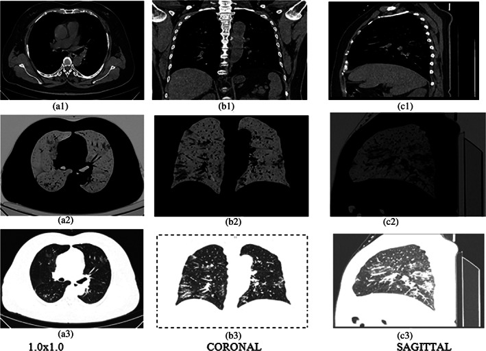
Three types of images (a) 1.0 × 1.0, (b) CORONAL, (c) SAGITTAL. In first row‐original; second row‐ROI, third row‐invert of the 61 years old patient

Three types of images (a) 1.0 × 1.0, (b) CORONAL, (c) SAGITTAL. In first row‐original; second row‐ROI, third row‐invert of the 61 years old patient