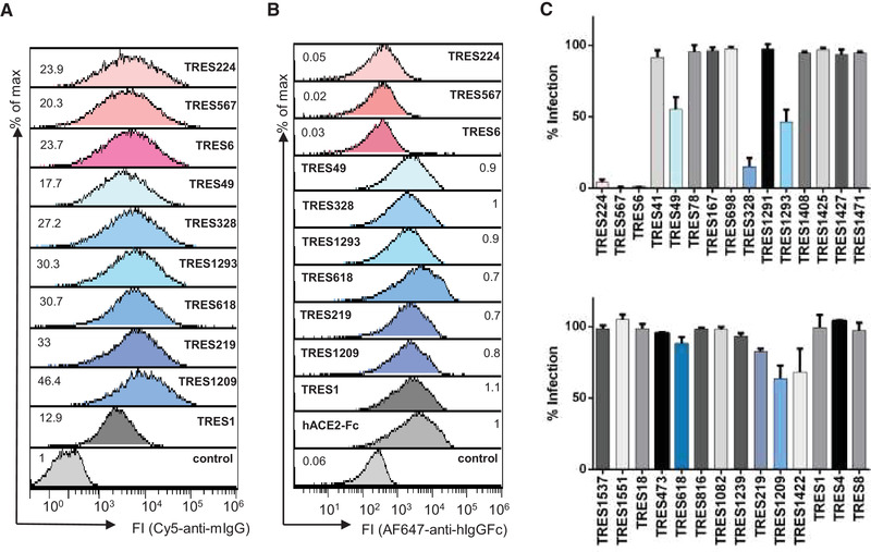
Figure 2.

Screening of hybridoma supernatants. (A) The binding of antibodies from undiluted TRES hybridoma supernatants to HEK‐293T cells expressing the SARS‐CoV‐2 spike protein was detected with a fluorescence‐conjugated murine pan IgG antibody. Numbers indicate the relative mean fluorescence intensity. (B) Detection of hACE2‐competing TRES antibodies. Numbers depict the relative mean fluorescence intensity. (C) Vero‐E6 cells were infected with the SARS‐CoV‐2 MUC‐IMB‐1 isolate in the presence or absence of undiluted TRES hybridoma supernatants. SARS‐CoV‐2 infection was quantified after 20 to 24 h by staining as described in Fig. 1F. The mean and standard deviation of triplicates of one experiment are shown.
