
Figure 4.
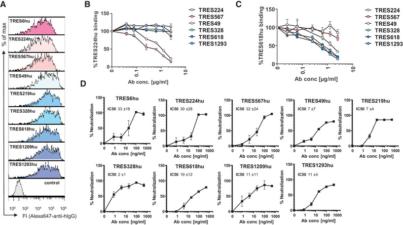
Characterization of recombinant human TRES antibodies. (A) Flow cytometric analysis of HEK‐293T cells expressing the SARS‐CoV‐2‐S protein and stained with recombinant humanized IgG1 TRES (TREShu) antibodies and a fluorochrome‐labeled secondary antibody against human IgG‐Fc. A non‐S binding human antibody served as a negative control. (B, C) HEK‐293T cells expressing the SARS‐CoV‐2 spike protein were incubated with recombinant TRES antibodies with a human Fcγ1 region and serially diluted TRES hybridoma antibodies with a murine Fcγ. Bound recombinant human TRES224hu (B) or TRES618hu (C) were detected with a mouse Alexa647‐labeled antibody directed against the human Fcγ region. The mean percentages of binding and SEM of one experiment performed in triplicates are shown. (D) The SARS‐CoV‐2 neutralizing activity of the human recombinant TRES antibodies was analyzed as described in Fig. 3B. Shown are means and SEM of triplicates of one representative experiment out of three. Also given are the mean and standard deviation of IC50s, given in ng/ml, of the three independent experiments, calculated as described in Fig. 3B