
Figure 2.
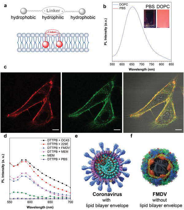
a) Schematic illustration of DTTPB structural characteristics and its interaction with membrane structure. b) PL spectra of DTTPB (5 µm) in PBS solution with or without 5 mg mL−1 DOPC. Inset: corresponding FL images of DTTPB in PBS solution with or without DOPC under a hand‐held UV lamp at 365 nm. c) Co‐localization imaging of HeLa cells stained with DTTPB (red) and CellMask Green (green). The 561 nm laser and 620–720 nm emission filter were used for DTTPB. The 488 nm laser and 510–550 nm emission filters were used for CellMask Green. Scale bar: 10 µm. d) PL spectra of DTTPB (5 µm) in different kinds of virus solutions, MEM media, or PBS solution. Excitation wavelength: 490 nm. e,f) Schematic illustrations of the structural characteristics of (e) human coronaviruses and (f) FMDV.