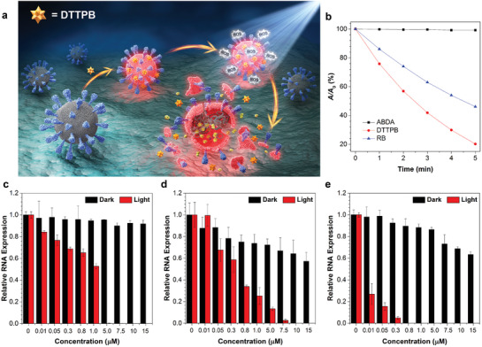
Figure 3.

a) Schematic illustration of the PDI process of coronaviruses with DTTPB. b) Decomposition rates of ABDA in the absence or presence of DTTPB or RB under light irradiation (20 mW cm−2), where A 0 and A are the initial and final absorbance of ABDA at 378 nm, respectively. c–e) qPCR studies of relative RNA expression of (c) FMDV, (d) HCoV‐OC43, and (e) HCoV‐229E. The viruses were treated with white‐light irradiation (9 mW cm−2) for 20 min, followed by infecting host cells and RNA extraction. Data were expressed as mean ± SE, number of duplicates: 3.
