
Figure 4.
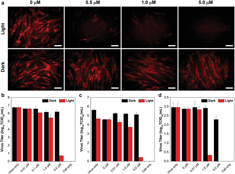
a) Immunofluorescence studies of MRC‐5 cells infected with HCoV‐OC43, which were pre‐treated with different concentrations of DTTPB and then irradiated with 9 mW cm−2 white light for 20 min. These MRC‐5 cells were then subjected to immunostaining with primary anti‐OC43 specific polyclonal antibody and TRITC Goat Anti‐Mouse IgG (H+L) secondary antibody, followed by imaging with a FL microscope (λex: 510–550 nm, λex: 570–750 nm). Scale bar: 50 µm. b–d) TCID50 assay for detecting the live viral (b) FMDV, (c) HCoV‐OC43, (d) and HCoV‐229E titers. The viruses were incubated with different concentrations of DTTPB with or without 9 mW cm−2 white‐light irradiation for 20 min. Data were expressed as mean ± SE, number of duplicates: 8.