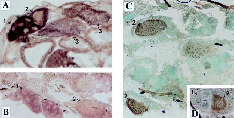
FIG. 4.
Expression of e(y)1 in different tissues of Oregon R flies. (A and B) In situ hybridization of a frontal tissue section of female abdomen with the DIG-labeled e(y)1 antisense (A) and sense (B) RNA probes. (C and D) Immunostaining of a frontal tissue section of female abdomen with antibodies to e(y)1 protein. Horseradish peroxidase and DAB were used for visualization; the tissue was counterstained with fast green. One can see a high level of e(y)1 transcription and expression in ovaries: 1, in trophocytes; 2, in primary oocytes; 3, in mature oocytes. Note that while the level of e(y)1 mRNA content is high in trophocytes and mature oocytes and low in primary oocytes, the TAFII40 protein is predominantly detected in oocytes rather than in trophocytes (C). Magnification, ×130.

