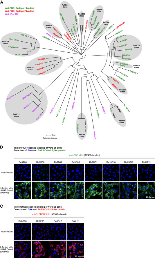
The sequences of the isolated VHH antibodies (49 anti‐RBD and 10 anti‐S1ΔRBD nanobodies) were aligned using Clustal Omega (Sievers
et al,
2011). The circular phylogram was reconstructed with Dendroscope (Huson & Scornavacca,
2012). Where applicable, sequence classes are indicated. The anti‐RBD nanobodies are colored according to the two RBD epitopes identified in Fig
5 (with epitope 1 in green, epitope 2 in red, and asterisks marking nanobodies included in Fig
5). Anti‐S1ΔRBD nanobodies are colored in magenta. VHH antibodies that did not neutralize SARS‐CoV‐2 are shown in parentheses. See main text and Fig
5 for details.

