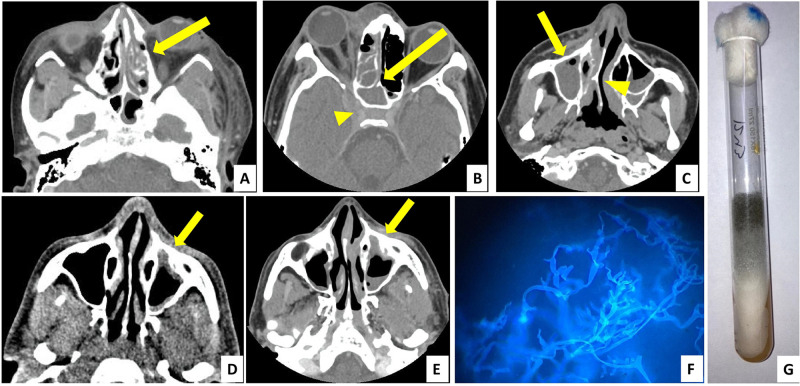
Figure 2.
Diagnosis of mucormycosis—radiology (A–E axial section, contrast-enhanced computed tomography images) and microbiology (F and G). (A) Left ethmoidal sinusitis with bony erosion (yellow arrow). (B) Right ethmoidal and sphenoid sinusitis (yellow arrow) with involvement of orbit and orbital apex, extension into cavernous sinus (yellow arrowhead). (C) Right maxillary sinusitis (yellow arrow) with deviated nasal septum (yellow arrowhead). (D) Left maxillary sinus showing thickened mucoperiosteal lining (yellow arrow). (E) Same patient from (D) showed increase in thickening and involvement of maxillary sinus after 1 week (yellow arrow). (F) Mucorales with aseptate hyphae seen on potassium hydroxide mount with calcofluor white stain. (G) Mucorales growth as greyish white colonies on Sabouraud dextrose agar medium.

