Figure 1.
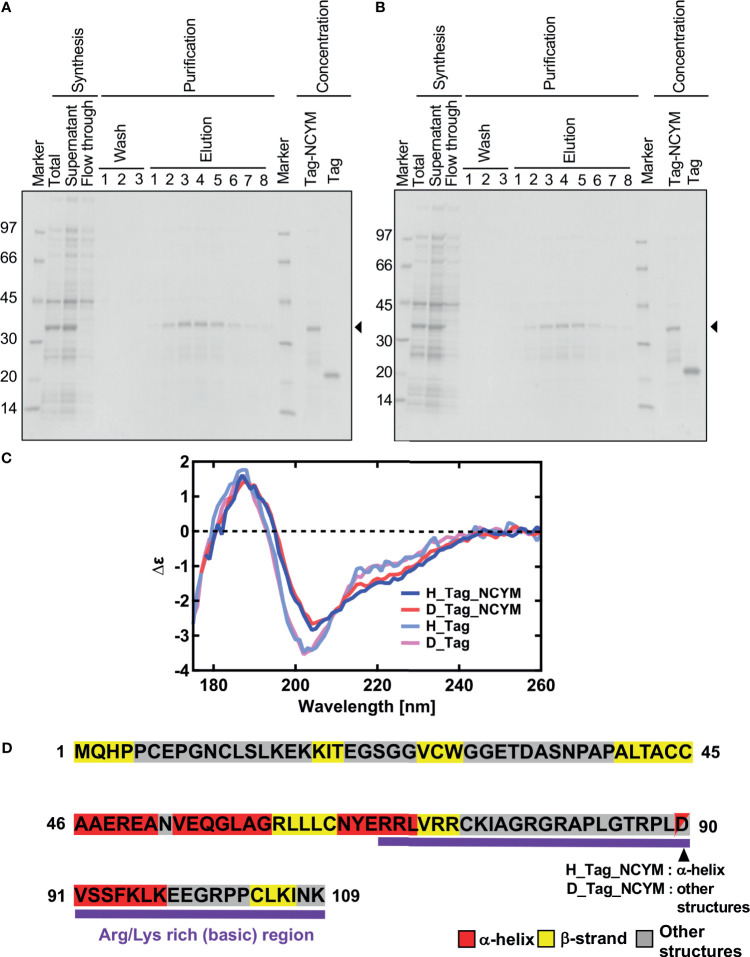
Vacuum-ultraviolet circular dichroism (VUVCD) analyses revealed the secondary structure of NCYM. (A) NCYM with SUMO tag (arrow) and the isolated SUMO tag were synthesized and purified using an in vitro cell-free system. (B) Perdeuterated NCYM with SUMO tag (arrow) and the isolated SUMO tag were synthesized and purified using an in vitro cell-free system. (C) VUVCD spectra for hydrogenated SUMO-tagged NCYM (H_Tag_NCYM), perdeuterated SUMO-tagged NCYM (D_Tag_NCYM), hydrogenated SUMO tag (H_Tag), and perdeuterated SUMO tag (D_Tag). (D) Secondary structure of NCYM predicted using the neural network. Secondary structures are highlighted in red, yellow, and gray for α-helix, β-strand, and other structures, respectively. Arg/Lys-rich (basic) region is highlighted in purple.

