The following was originally published in Volume 25, Number 1, pages 43–53 (doi: https://doi.org/10.3727/096504016X14719078133285). In the original article there were some inappropriately organized images of Western blots and some images did not show the best experimental results. We have revised the figures to correct these images (Figs. 2A, 4A, 6A, 7B and C, and 8C and D). The figures have been reorganized using images from repeated experiments to rectify the errors. Corrected versions of the figures are shown here and the figures have been replaced with the corrected versions in the original published article in the online site (https://www.ingentaconnect.com/contentone/cog/or/2017/00000025/00000001/art00006). The corrections do not change any results or conclusion of the article. The authors apologize for any inconvenience caused.
Abstract
The role of miRNAs in the radiosensitivity of glioma cells and the underlying mechanism is still far from clear. In the present study, we detected six downregulated and seven upregulated miRNAs in the serum after radiotherapy compared with paired serum samples before radiotherapy via miRNA panel PCR. Among these, miR-17-5p was highly reduced (fold change = −4.21). Further, we validated the levels of miR-17-5p in all serum samples with qRT-PCR. In addition, statistical analysis suggested that a reduced miR-17-5P level was positively associated with advanced clinical stage of glioma, incidence of relapse, and tumor differentiation. Moreover, we provided evidence that irradiation markedly activated autophagy and decreased miR-17-5p in the glioma cell line. Further, we demonstrated that irradiation-induced autophagy activation was mediated by beclin-1, and downregulation of beclin-1 via siRNA significantly abolished the irradiation-activated autophagy. Interestingly, we demonstrated that miR-17-5p could directly target beclin-1 via luciferase gene reporter assay. Exotic expression of miRNA-17-5p decreased autophagy activity in vitro. In nude mice, miRNA-17-5p upregulation sensitized the xenograft tumor to irradiation and suppressed irradiation-induced autophagy. Finally, pharmacal inhibition of autophagy markedly enhanced the cytotoxicity of irradiation in RR-U87 cells.
Key words: Glioma, MicroRNA-17-5p, Beclin-1, Autophagy, Radiosensitivity
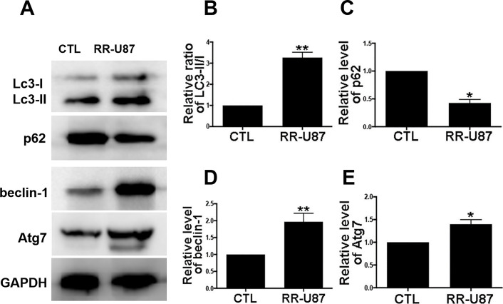
Figure 2.
Irradiation-activated autophagy in U87 cells. (A) Autophagy activity was detected by Western blot for LC3-I/II, p62, beclin-1, and Atg7. (B–E) Relative levels of ratios of LC3-II/I (B), p62 (C), beclin-1 (D), and Atg7 (E) were measured using ImageJ and normalized to GAPDH. CTL (control) denotes U87 cells without irradiation, and RR-U87 denotes radioresistant U87 cells. Data are presented as mean ± SD. *p < 0.05 and **p < 0.01 compared with control group.
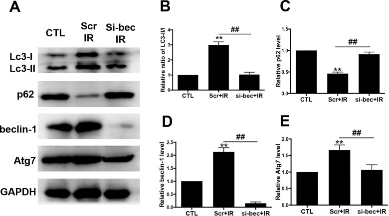
Figure 4.
Irradiation-induced autophagy activation was mediated by beclin-1. (A) U87 cells were transfected with siRNA for beclin-1 (si-bec) for 24 h and then exposed to irradiation (2 Gy/min). Levels of LC-3I/II, p62, beclin-1, and Atg7 were detected with Western blot. (B–E) Relative levels of ratios of LC3-II/I (B), p62 (C), beclin-1 (D), and Atg7 (E) were measured using ImageJ and normalized to GAPDH. CTL (control) denotes U87 cells without irradiation. Scr denotes scramble. Si-bec denotes siRNA for beclin-1. IR denotes irradiation. Data are presented as mean ± SD from at least three independent experiments. **p < 0.01 compared with control group and ##p < 0.01 compared with the indicated groups.
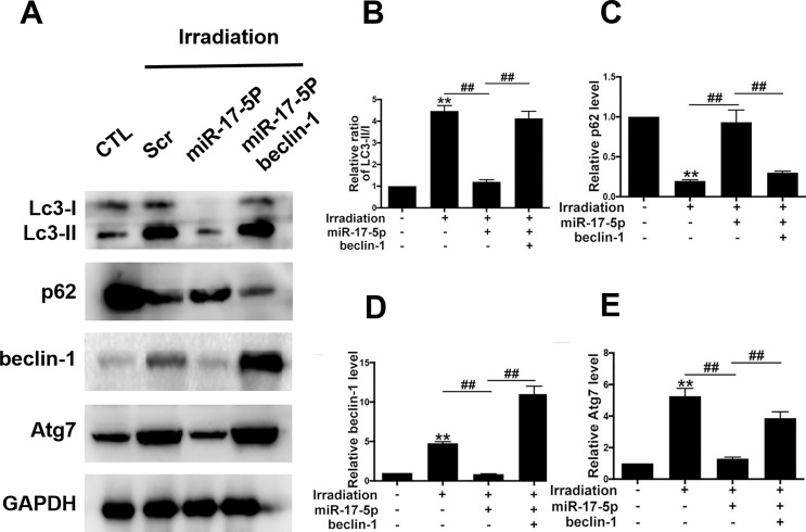
Figure 6.
Exotic expression of miR-17-5p decreased autophagy activity via targeting beclin-1. (A) U87 cells were transfected with miR-17-5p mimics with or without beclin-1 overexpression plasmids. Then the cells were subjected to irradiation, and the autophagy activity was tested by Western blot. (B–E) Relative levels of ratio of LC3-II/I (B), p62 (C), beclin-1 (D), and Atg7 (E) were measured using ImageJ and normalized to GAPDH. Data are presented as mean ± SD from at least three independent experiments. **p < 0.01 compared with control group and ##p < 0.01 compared with the indicated groups.
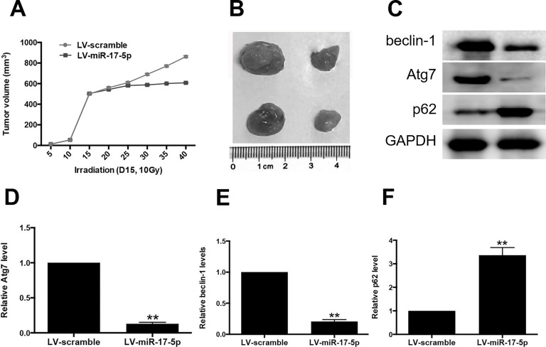
Figure 7.
miR-17-5p sensitized xenograft tumor to irradiation and inhibited irradiation-induced autophagy. (A) Nude mice were subcutaneously injected with U87 cells infected with control lentivirus vector or miR-129-5p overexpression lentivirus vector. The tumors were irradiated (10 Gy) on day 15 when the average tumor volume reached about 500 mm3. Tumor volume was monitored over time as indicated. (B) The tumors were excised on day 40. miR-129-5p overexpression caused a significant decrease in tumor volume. (C–F) the autophagy activity was determined by Western blot for beclin-1, Atg7, and p62. Relative density of protein bands was measured using ImageJ and normalized to GAPDH. Data are presented as mean ± SD from at least three independent experiments. **p < 0.01 compared with LV-scramble group.
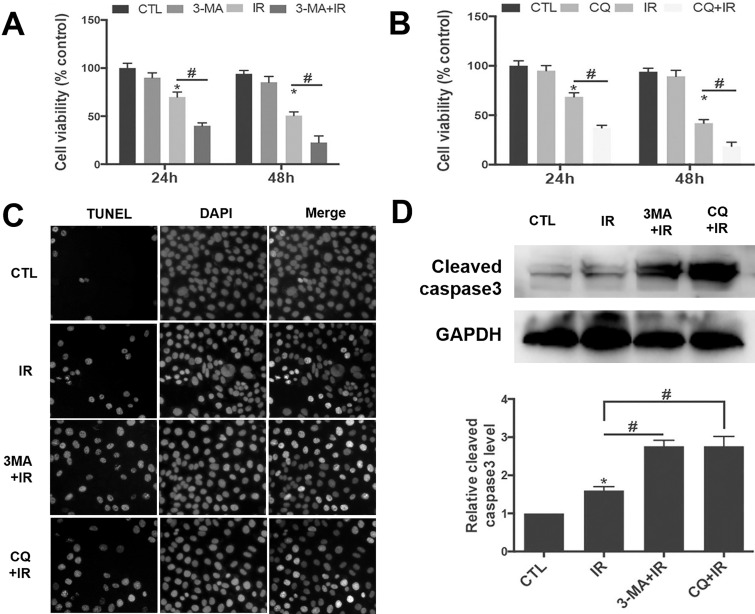
Figure 8.
Pharmacal inhibition of autophagy sensitized RR-U87 cells to irradiation. (A, B) RR-U87 cells were pretreated with 4 μM 3-MA or 20 μM CQ for 1 h and then exposed to irradiation (2 Gy/min). The cell viability at 24 and 48 h after irradiation was measured by MTT. Data are presented as percentage normalized to control group and are presented as mean ± SD from at least three independent experiments. CTL, control; IR, irradiation. Compared with the control group, 3-MA or CQ pretreatment significantly sensitized RR-U87 cells to irradiation-induced cell death. (C) TUNEL assay was conducted to detect the apoptosis in RR-U87 cells after autophagy was suppressed with 3-MA or CQ. Scale bar: 50 μm. (D) Cleaved caspase 3 was detected with Western blot, and relative level of cleaved caspase 3 was measured using ImageJ and normalized to GAPDH. Data are presented as mean ± SD from at least three independent experiments. *p < 0.05 compared with control group and #p < 0.05 compared with the indicated groups.


