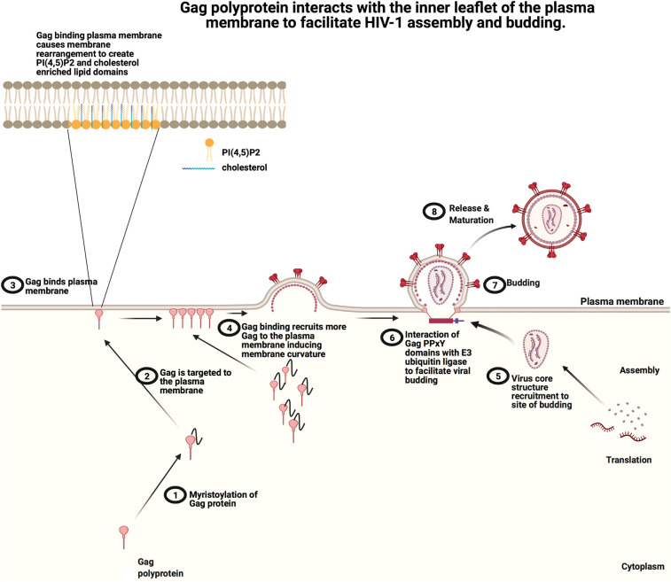
Figure 2. Schematic of the HIV-1 Gag assembly and budding from the host cell plasma membrane.
(1) The Gag polyprotein is myristoylated on a N-terminal glycine, which (2) facilitates membrane insertion into the plasma membrane inner leaflet. (3) At the plasma membrane, Gag is able to interact with PI(4,5)P2 and PS, which helps to facilitate Gag oligomerization and cluster PI(4,5)P2 and cholesterol. (4) Gag oligomerization and clustering of PI(4,5)P2 and cholesterol into enriched regions of the plasma membrane, further induces recruitment of Gag proteins and likely facilitates changes in membrane curvature. (5) As the Gag mediated bud site matures, the virus core is recruited and (6) interactions with host proteins such as an E3 ligase (7) facilitate viral budding. (8) Finally, a mature HIV Gag virion is released from the plasma membrane. Adapted from ‘HIV Replication Cycle’ by BioRender.com (2021). Retrieved from https://app.biorender.com/biorender-templates.

