Fig. 7.

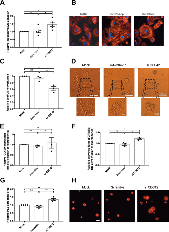
CDC42 mediated the effect of miR-204-5p on PLS. ( A ) CDC42 knock-down increased the adhesion of megakaryocytes in a fibrinogen-coated channel in dynamic conditions and ( B ) led to an actin cytoskeleton organization similar to that observed after miR-204-5p transfection with an increase in the stress-fiber content. Images were taken using confocal microscopy of megakaryocytes, with polylobed nucleus labeled with DAPI ( blue ) and F-actin labeled with TRITC-phalloidin ( red ) (scale bars = 20 μm). ( C ) Knock-down of CDC42 decreased the relative proPLS network area and ( D ) displayed a similar pattern to that observed after miR-204-5p transfection (Si-3000 camera, scale bars = 100 μm). ( E ) CDC42 silencing did not impact CD62P expression and ( F ) increased relative GPIIbIIIa expression in activated PLS and ( G and H ) enhanced the PLS spreading area on fibrinogen-coated coverslips (LSM700 microscope, scale bars = 20 μm). Results are expressed relative to the mock condition. n = 3 to 5 independent experiments; * p < 0.05, ** p < 0.01. PLS, platelet-like structure.
