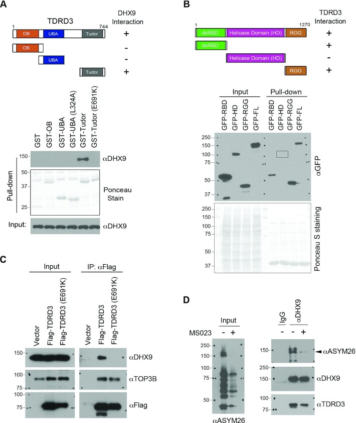
Figure 2.
Characterization of the interaction between DHX9 and TDRD3. (A) Mapping the region of TDRD3 that interacts with DHX9. GST-tagged TDRD3 truncation constructs were generated, containing the oligonucleotide/oligosaccharide-binding (OB)-fold, the ubiquitin-associated domain (UBA; wild type or with L324A mutation), and the Tudor domain (Tudor; wild type or with E691K mutation), respectively. A graphic summary of their interactions with DHX9 is shown. A GST pull-down assay was performed by incubating the recombinant GST-fusion proteins with MCF7 cell lysates. The pull-down samples were detected by western blot analysis using an αDHX9 antibody. The GST-fusion proteins were visualized by Ponceau staining. (B) Mapping the region of DHX9 that interacts with TDRD3. GFP-tagged full-length or truncations of DHX9 constructs were generated. The locations of the N-terminal double-stranded RNA binding domain (dsRBD), the helicase domain (HD), and the C-terminal RGG-containing domain (RGG) are indicated (upper panel). A graphic summary of their interactions with TDRD3 is shown. A GST pull-down assay was performed by incubating the recombinant GST-Tudor with MCF7 lysates that were transfected with different GFP-DHX9 fusion vectors. Both the input and pull-down samples were detected by western blot analysis using an αGFP antibody. The GST-Tudor recombinant protein used in the binding was visualized by Ponceau staining. (C) The interaction of TDRD3 with DHX9 requires a functional Tudor domain. A co-IP assay was performed to assess the interaction of Flag-tagged WT and methylarginine binding-deficient (E691K) TDRD3 with endogenous DHX9 in MCF7 cells. The input and αFlag antibody-immunoprecipitated samples were analyzed by western blot using indicated antibodies. (D) DHX9 interacts with TDRD3 in an arginine methylation-dependent manner. The interaction of DHX9 with TDRD3 was detected by co-IP in MCF7 cells treated with vehicle (−) or with the type I PRMT inhibitor MS023 (+). The level of cellular ADMA was detected by western blot using a pan-ADMA antibody (ASYM26). The DHX9-immunoprecipitated samples were probed for methylation and interactions with TDRD3 using the ASYM26 and αTDRD3 antibodies.

