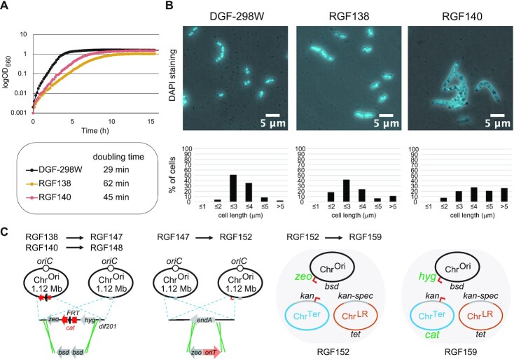
Figure 4.
Phenotypes of tripartite-genome strains. (A) Growth of tripartite-genome strains. Cells were incubated at 37°C with shaking (60 rpm) in antibiotic-free LB media using L-shaped tubes. Doubling times were computed from the growth curve data. (B) Overlay of phase contrast and DAPI stained microscopy images of tripartite-genome strains (upper panels) and distribution of their cell lengths (lower panels). Exponential phase cells growing at 37°C in antibiotic-free LB media were observed and counted. The distribution of cell lengths from 210 DGF-298W cells, 239 RGF138 cells, and 203 RGF140 cells are shown below each picture. (C) A diagram depicting the genetic modifications to RGF138 and RGF140.

