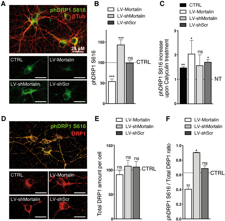
Figure 5.
Mortalin impacts mitochondrial morphology by modulating Drp1-S616 phosphorylation. Rat embryonic primary cortical neurons were transduced on DIV 3 with lentiviral vectors (LV) to induce over- (LV-Mortalin) or under- (LV-shMortalin, clone A) expression of Mortalin. A lentiviral vector expressing a shRNA with a scramble sequence was used as a control (LV-shScr). Neurons were then fixed on DIV 12 and the active form of Drp1, phosphorylated on serine 616, was revealed by pDrp1-S616 staining. (A–C) The neuronal network was visualized with the neuronal marker βIII-tubulin (βIII-Tub). On each randomly taken picture (A), the total amount of pDrp1 was measured (integrated staining density, Image J software) and normalized on the βIII-tubulin staining area. For each experiment, 4 pictures were taken per condition and the corresponding mean ratios were normalized relative to control, non-transduced neurons. (B). The same was done upon treating neurons with Calyculin-A for 30 min before fixation, in order to inhibit phosphatase activity (C). The results are presented as means ± SD of 4 experiments. (D–F) Neurons were co-strained with antibodies directed against total Drp1 and pDrp1-S616 (D). For each randomly taken picture, we evaluated the total and phosphorylated protein amounts in βIII-Tub + cells, using the integrated staining density (Image J software). The values obtained for total Drp1 were normalized on the signal for non-transduced neurons in each experiment and are presented as means ± SD of 3 independent experiments (E). Ratios of the integrated staining densities for pDrp1-S616 on total Drp1 were also measured and normalized on the ratio obtained for un-transduced cells and presented as means ± SD of 3 independent experiments (F). **p < 0.01, by Mann–Whitney non parametric test.

