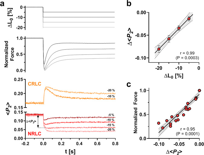
Fig. 3. Orientation changes of the NRLC and CRLC probe in response to shortening of Ca2+-activated muscle.
a Upper panel: muscle length change (ΔL0); middle panel: normalized force; bottom panel: <P2> from the NRLC (red) and CRLC probe (orange). b Length-dependence of the change in <P2> (Δ<P2>) from NRLC measured at the end of the <P2> transient. Data are shown as means ± s.e.m. (n = 5 for −20% ΔL0 and −10% ΔL0; and n = 4 for −5% ΔL0 and −15% ΔL0). c Plot of steady state <P2> change (Δ<P2>) of NRLC versus normalized isometric force (n = 23; pooled data from n = 5 trabeculae). Solid line represents linear regression with 95% confidence interval indicated by dotted lines. Pearson correlation coefficient (r) and p value for a two-tailed student’s t test are shown on the bottom right. Source data are provided as a Source Data file.

