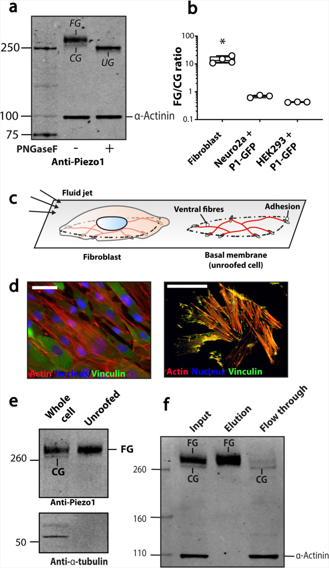
Fig. 2. N-glycosylation of endogenous Piezo1 in human fibroblasts.
a A representative western blot of untreated immortalised human foreskin fibroblasts versus PNGaseF treated fibroblast lysate probed using the anti-Piezo1 antibody and anti-α-actinin antibody as a loading control. b The ratio [FG/CG] of the upper band (FG) and lower band (CG) of fibroblast Piezo1 and Piezo1 heterologously expressed in Neuro2A and HEK293T. c Schematic illustration of an intact fibroblast, and an unroofed fibroblast. d A representative image of an intact fibroblast (left panel), and an unroofed fibroblast (right panel) using standard wide-field microscopy and a 63× oil objective. e A representative western blot of intact fibroblast lysate versus unroofed fibroblast lysate probed using anti-Piezo1 antibody and anti-α-tubulin antibody as a loading control to confirm unroofing. f A representative western blot of biotinylated immortalised human foreskin fibroblasts. (CG core-glycosylated, FG fully glycosylated) *p < 0.05 determined by Kruskal–Wallis test with Dunn’s post-hoc test.

