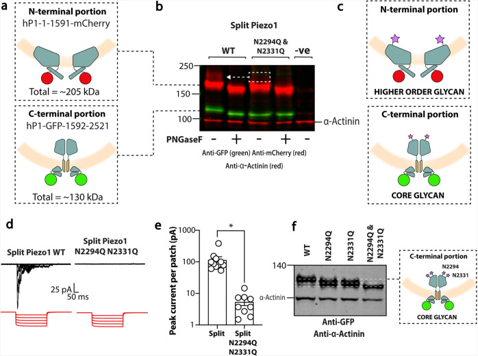
Fig. 5. N-glycosylation of Piezo1 in a split construct.
a Diagram depicting the human split Piezo1 protein. b Representative blot showing the split human Piezo1 protein and the split human Piezo1 protein with the double cap mutant N2294Q and N2331Q with and without PNGaseF treatment. c Diagram illustrating where glycans are likely to be located. d Electrophysiological recordings of Piezo1−/− HEK293T cells expressing human split Piezo1 and the double cap mutant N2294Q and N2331Q in the cell-attached configuration in response to negative pressure applied using a high-speed pressure-clamp (red). e Quantification of peak current elicited per patch. f Representative blot showing the C-terminal portion of the split human Piezo1 protein and the mutants N2294Q, N2331Q and N2294Q/N2331Q. Note that the single mutants run at the same size but the double mutant is smaller. *p < 0.05 determined by Mann–Whitney U-test. −ve represents an un-transfected control.

