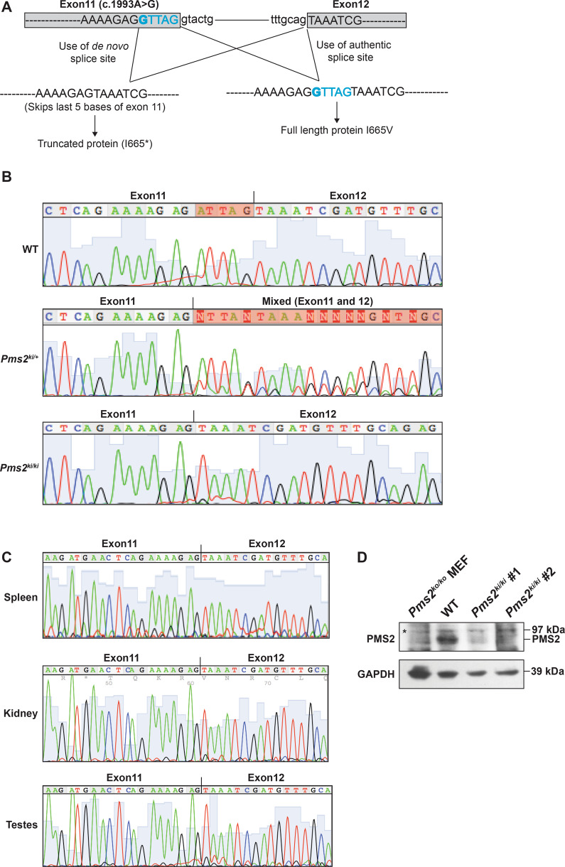
Fig. 1. Pms2ki/ki mice exhibit skipping of five bases of exon 11.
A Schematic representation of splicing pattern due to c.1993A>G mutation. Relevant exons are shown in box and intron as straight line. Exon sequences are in capital letters and intron sequences are in small letter. The mutation is in bold and the five base of exon 11 that are deleted in the transcript due to generation of new splice site are in blue. B Chromatogram showing the sequence of Pms2 transcript from colon of wild type (WT), Pms2ki/+ and Pms2ki/ki mice. Highlighted bases in WT chromatogram shows the region skipped due to c.1993A>G mutation. Transcript sequence from heterozygous mice showing mixed sequence is highlighted. C Pms2 transcript sequences from different tissues (spleen, kidney and testes) of Pms2ki/ki mice showing the deletion of last five bases of exon 11. D Expression of PMS2 protein in wild-type (WT) and Pms2ki/ki mice. Proteins were isolated from spleen of mice with indicated genotypes. Two independent Pms2ki/ki mice were used and they are numbered as #1 and #2. Proteins isolated from immortalized mouse embryonic fibroblast of Pms2ko/ko (null) mice (127TAg) [ATCC; cat# CRL-2817TM] was used as negative control. GAPDH was used as loading control. Asterisk designates non-specific band. Protein size markers are indicated on right side.

