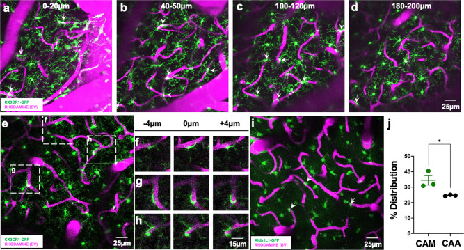
Fig. 1. Ramified CX3CR1+ myeloid cells associate with brain capillaries.
a–d Representative 20-µm-thick two-photon projection images from a CX3CR1GFP/+ adult brain showing myeloid cells (green) and the vasculature (rhodamine in magenta) at varying tissue depths between the brain surface and 200 µm of the cortex. Arrows identify capillary-associated ramified myeloid cells. e Representative 20-µm-thick in vivo two-photon image from a CX3CR1GFP/+ adult brain showing ramified myeloid cells (green) and the vasculature (rhodamine in magenta) in the cortex. f–h Representative images from boxed regions in (e) showing somal interactions between the ramified myeloid cells and capillaries in an 8 µm tissue volume. i, j Representative 20-µm-thick two-photon projection image (i) and quantification (j) from an ALDH1L1GFP/+ P30 brain showing astrocytes (green) and the vasculature (rhodamine in magenta) in the cortex. Capillary-associated astrocytes (CAAs, arrows in i) density is compared to capillary-associated myeloid (CAM) density. n = 3 mice each. Representative images in (a–h) were observed in five mice and in (i) was observed in three mice. Data are presented as mean values ± SEM. *p < 0.05. Two-sided unpaired Student’s t test.

