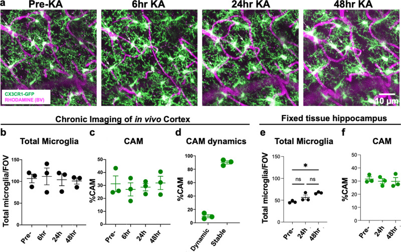
Fig. 5. CAM interactions are not altered by increased neuronal activity.
a Representative in vivo two-photon projection images from the same field of view in a CX3CR1GFP/+ adult brain showing capillary- (magenta) associated microglia (green) before and up to 48 h after KA-induced seizures. b, c Quantification of total microglial (b) and capillary-associated microglial (c) density over time before and following seizures. d Quantification of the stable and dynamic CAMs following seizures. e–f Quantification of total microglial (e) and capillary-associated microglial (f) density from fixed slices in the hippocampus following seizures. n = 3 mice each. Data are presented as mean values ± SEM. *p < 0.05. n.s. not significant. Two-sided unpaired Student’s t test in b, c, e, and f.

