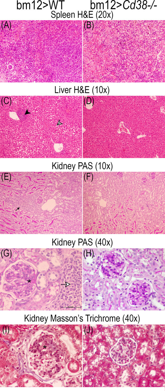
Figure 11.

Representative microphotographs of the histological studies performed on the spleen, liver, and kidney 3-μm sections at 8 weeks of the adoptive transfer of spleen cells from female bm12 to female WT (left panels) or Cd38−/− (right panels) mice. (A, B) Spleen, (C, D) liver, and (E–J) kidney. (A–D) Spleen and liver tissue sections were stained with H&E. (E–H) Kidney tissue sections were stained with PAS. (I, J) Kidney sections were stained with Masson’s trichrome. (A, B) ×20 magnification; (C–F) ×10 magnification; (G–J) ×40 magnification. (G) Scale bar: 50 μm. Note in the liver from WT cGVHD mice the presence of mild perivascular chronic inflammatory infiltrate [black head arrow in (C)] and macrovesicular steatosis [black and white head arrow in (C)]. Note in the kidney of WT cGVHD the periglomerular chronic inflammatory infiltrate [small black arrow in (E)] and the increased glomerular size [asterisks in (G) and (I)].
