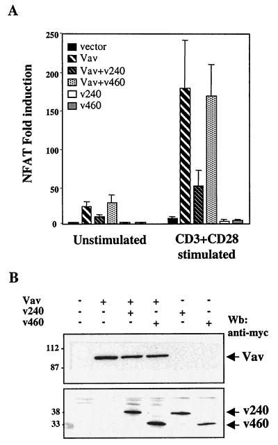
FIG. 5.
Vav-mediated NFAT activation is inhibited by hSiah2. (A) T-Ag Jurkat cells (107) were transfected with NFAT and pSVβ-galactosidase reporter plasmids (5 and 1 μg, respectively) and 20 μg of either an empty vector (vector), pEF-Vav (Vav), pCAN-v240 (v240), pCAN-v460 (v460), or a combination of the vectors as indicated. A total of 106 cells were either left unstimulated or stimulated after 24 h with anti-CD3 plus anti-CD28 for 8 h. Luciferase activity was measured and corrected for β-galactosidase activity, and the results were expressed as average fold induction relative to unstimulated cells transfected with the empty vector. The data are representative of four independent experiments. The basal activity and the maximum NFAT responses were approximately 600 and 2 × 10⁵ AU, respectively. (B) T-Ag Jurkat cell lysates from panel A were analyzed by immunoblotting for expression of Vav and hSiah2 (v240 and v460). Wb, Western blot.

