Figure 1.
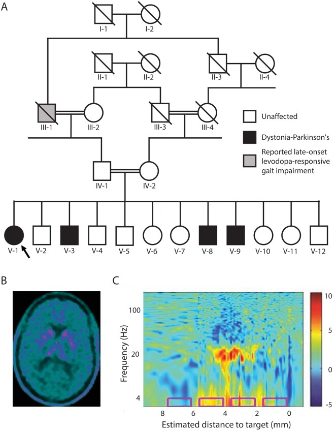
Clinical characteristics (A) Family tree of the studied family. Proband is indicated by an arrow. (B) Brain 18F-l-3,4-dihydroxyphenylalanine (18F-DOPA) PET/CT study (patient V-1) demonstrating severely impaired pre-synaptic striatal dopaminergic integrity. (C) Power spectral density plot demonstrates increased β oscillatory activity (around 20 Hz) in the right subthalamic trajectory of patient V-3, as a function of distance along the surgical trajectory. Estimated distance to target was preoperatively defined based on MRI. The colour scale represents log10 of power spectral density/average power spectral density. The implanted final contact depths of the permanent electrode are depicted by the pink rectangles.

